Dr Aleks Ponjavic
- Position
- University Academic Fellow
- Areas of expertise
- Single-molecule fluorescence imaging; light-sheet microscopy; super-resolution microscopy; high-speed fluorescence microscopy; cellular biophysics
- Location
- 102D, Sir William Henry Bragg Building
- Faculty
- Engineering and Physical Sciences
- School
- Physics and Astronomy/Food Science and Nutrition
Introduction
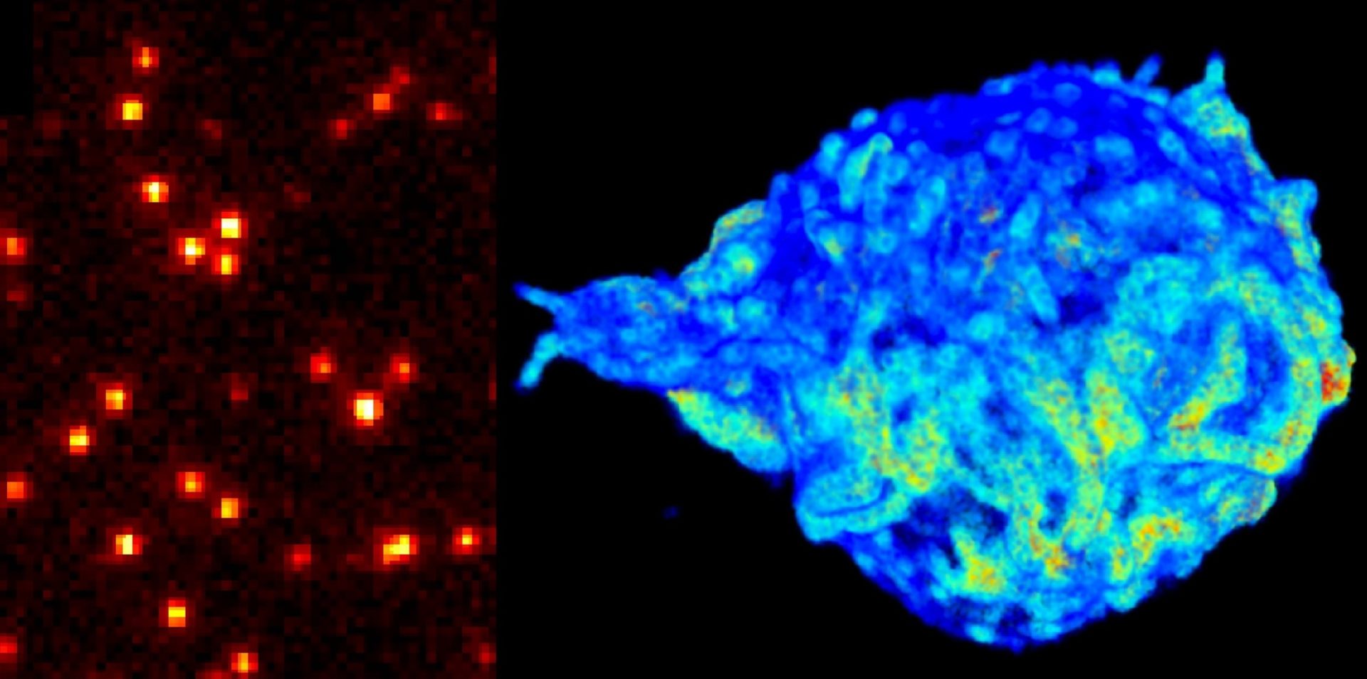
Cells make our bodies work through a myriad of complex processes that are generally poorly understood, even to this date, and our research focuses on improving the tools we have to visualise these processes. More specifically, our group uses fluorescence microscopy to observe the behaviour of the individual proteins that make up one the most basic components of cells.
Detailed research programme
Single-molecule fluorescence imaging
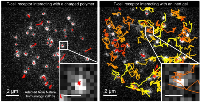
By using specialised microscopes and efficient laser excitation, it is possible to take videos of individual molecules as they move around and interact with their environment. We apply this technology to tackle fundamental biological questions such as “How do our immune cells identify germs?” and “How does cellular DNA change in shape to control gene expression?”.
Light-sheet microscopy
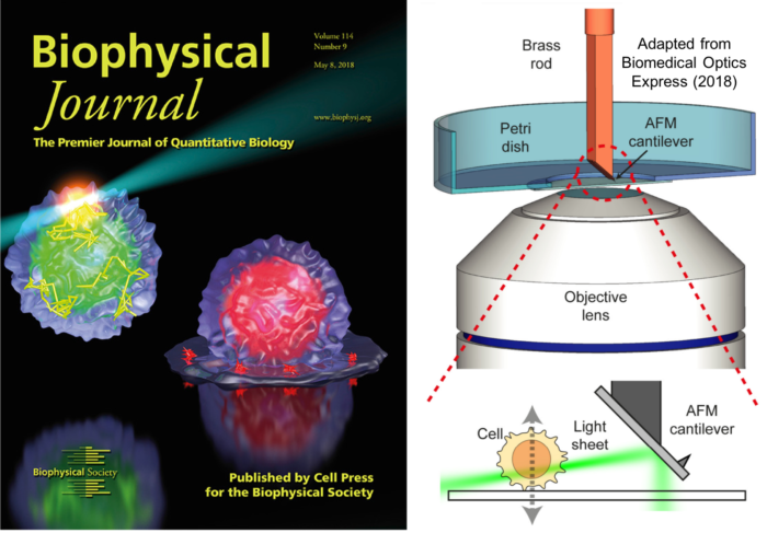
Traditional single-molecule imaging has mostly been limited to the glass-liquid interface as this enables extremely efficient laser excitation of molecules. By creating a thin sheet of light, it becomes possible to perform single-molecule imaging above the glass and inside cells. Our research focuses on implementing new ways of creating these light sheets to enable single-molecule studies in cells under more physiological conditions.
Nanoscale imaging
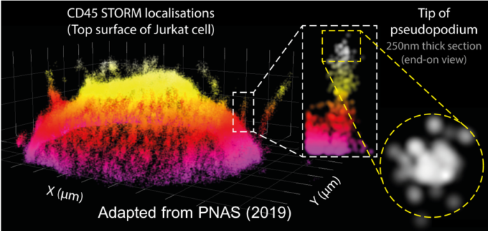
Super-resolution microscopy has been developed to overcome the diffraction limit of visible light (~250nm), which can be used to visualise structures down to the scale of the proteins themselves (~5nm). We develop and improve single-molecule super-resolution microscopy technology to investigate how proteins organise in 3D on the complex structures of cells.


