Dr Takashi Ochi
- Position
- University Academic Fellow
- Areas of expertise
- Cilia; Microtubule; Structural protein; Structural biology
- T.Ochi@leeds.ac.uk
- Location
- 10.127a Garstang
- Faculty
- Biological Sciences
- School
- Molecular and Cellular Biology

Introduction
The centrosome is a major microtubule-organising centre and probably the largest protein assembly having a few hundred-nanometre dimensions and found in animal cells. A centrosome is comprised of a pair of centrioles surrounded by pericentriolar material and centriolar satellites. The centriole has a characteristic 9-fold rotational symmetry, which is created by nine copies of parallelly aligned-microtubule triplets. Centrioles are also essential for generating cilia. Cilia have further extended-microtubule blades from centrioles, which are called basal bodies at cilia. Cilia can be largely classified into motile and non-motile: Motile cilia, which can be found more than one per cell, generated fluid flow in our airway, reproduction system & brain and locomotion for sperm cells. Many unicellular organisms such as green algae also use cilia to swim in fresh water. Non-motile cilia (primary cilia) are present one per cell and mediate intra-cellular signal transductions. Dysfunction of these organelles causes a wide range of diseases including primary microcephaly and ciliopathies. Also, abnormal structures of centrosomes such as centrosome amplification are often observed in cancer cells. The goal of my research is to understand how centrosomes and cilia maintain cellular homeostasis by determining the structures of the organelles using cryo-EM and X-ray crystallography.
Current major projects
- Biogenesis of accessory structures of the centriole
- Molecular mechanism of stabilising the structure of the centriole
- Understanding functions of centrosomal proteins at the atomic level
- Evolution of XRCC4/SAS6 superfamily in DNA repair and centrosomes
Detailed research programme
Structural studies of the centrosome to understand its protein organisation
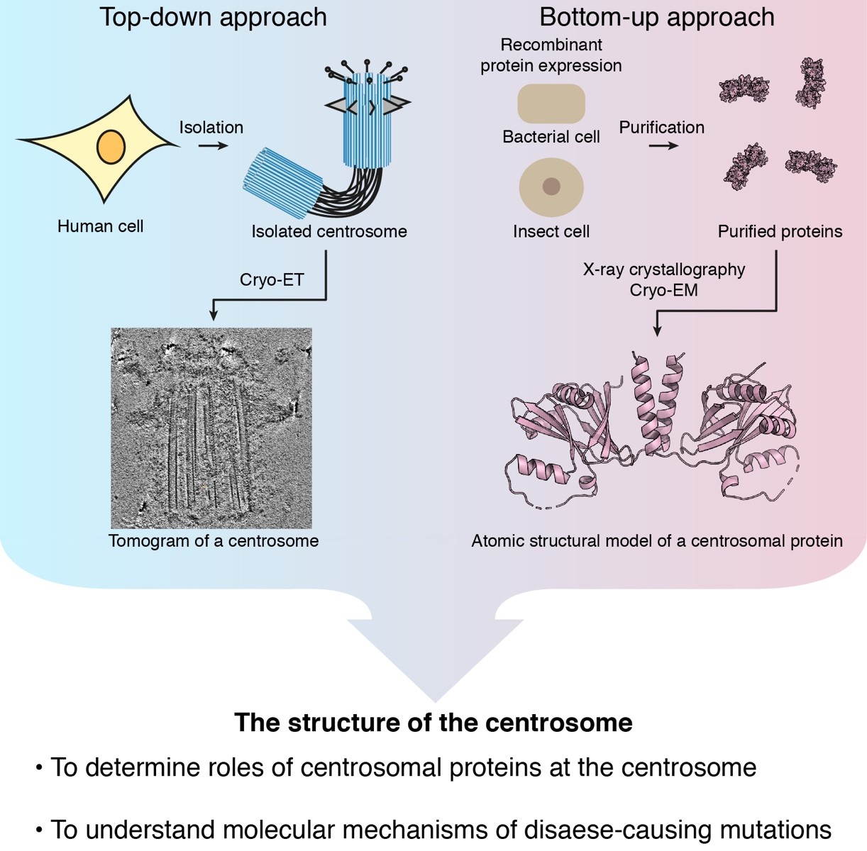
Centrosomes are considered to be stable and can be isolated from cells. A number of studies showed structural details of centrosomes using electron and light microscopies. However, we are still far from completely understanding how proteins are assembled to build centrosomes. To resolve this problem, we purify centrosomes and basal bodies from mammalian cells and observe structures of the organelles using cryo-electron tomography. Tomographic reconstruction of centrosomes and basal bodies show that common as well as unique structure features found among different cell types. It is our future challenge to reveal how cell / tissue specific structures are built on the common basis structure of centrosomes and basal bodies, and what functions of unique structures are.
Structural studies of centrosomal proteins to identify their functional residues
We also study the structure of each centrosomal protein to understand how the protein contributes to the structure of centrosomes. The main objective is to identify functional residues of proteins by determining structures of proteins. Since a protein tends to have more than one function, the knockout of a gene that removes multiple functions of a protein sometimes results in complex phenotypes. Protein structures are useful to separate these functions and study them individually in cells. We express recombinant proteins in bacterial or insect cells and use purified proteins to determine their structures using X-ray crystallography and cryo-EM. Identified functional residues are verified by making mutant constructs by site-directed mutagenesis and by testing their functions in vitro and in vivo.