Professor Michelle Peckham
- Position
- Professor of Cell Biology
- Areas of expertise
- Myosin; Actin; Cytoskeleton; Muscle; super-resolution imaging
- [email protected]
- Phone
- +44(0)113 343 4348
- Location
- 8.106 Astbury
- Faculty
- Biological Sciences
- School
- Molecular and Cellular Biology
Introduction
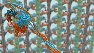
The molecular motor, myosin, is a key protein responsible for muscle contraction and movement in almost every eukaryotic cell. Our current research focuses on these and related cytoskeletal proteins. We research into how their activity is regulated, how they carry out their cellular functions, and effects of disease mutations. We use a wide range of tools, from characterisation of expressed and purified proteins to cultured muscle cells, super-resolution imaging (iSIM, STED, PALM and STORM), the use of Adhirons to label specific proteins in super-resolution imaging and to detect specific protein conformations. We use negative stain, Cryo-EM, and CryoET to investigate myosin structure as isolated molecules and ‘in situ’. The picture above shows our structure of shutdown smooth muscle myosin obtained by CryoEM (Nature 2020). We recently solved the structure of shutdown platelet myosin (non-muscle myosin 2A; Science Advances 2026).
Current major projects
- All aspects of molecular motors, with a focus on myosin, from structure, function, regulation, cellular organisation to effects of disease (Wellcome Trust Investigator Award)
- Development of super-resolution imaging approaches
Detailed research programme
Cytoskeleton
My lab has a broad interest in the cytoskeleton (actin filaments and microtubules). In skeletal and cardiac muscle, actin and myosin are organised into filaments with precise lengths to power contraction. In non-muscle cells, the actin cytoskeleton is much less well organized, is more dynamic, and these cells contain many different types of myosins, to enable dynamic cell behaviour. The contrast between the organisation and regulation of the cytoskeleton between highly ordered striated muscle, and crawling cells is fascinating. Mutations, or changes to expression levels are important drivers of a wide range of diseases from heart disease to bleeding disorders, and cancer.
Myosin: structure, function, regulation and roles in disease
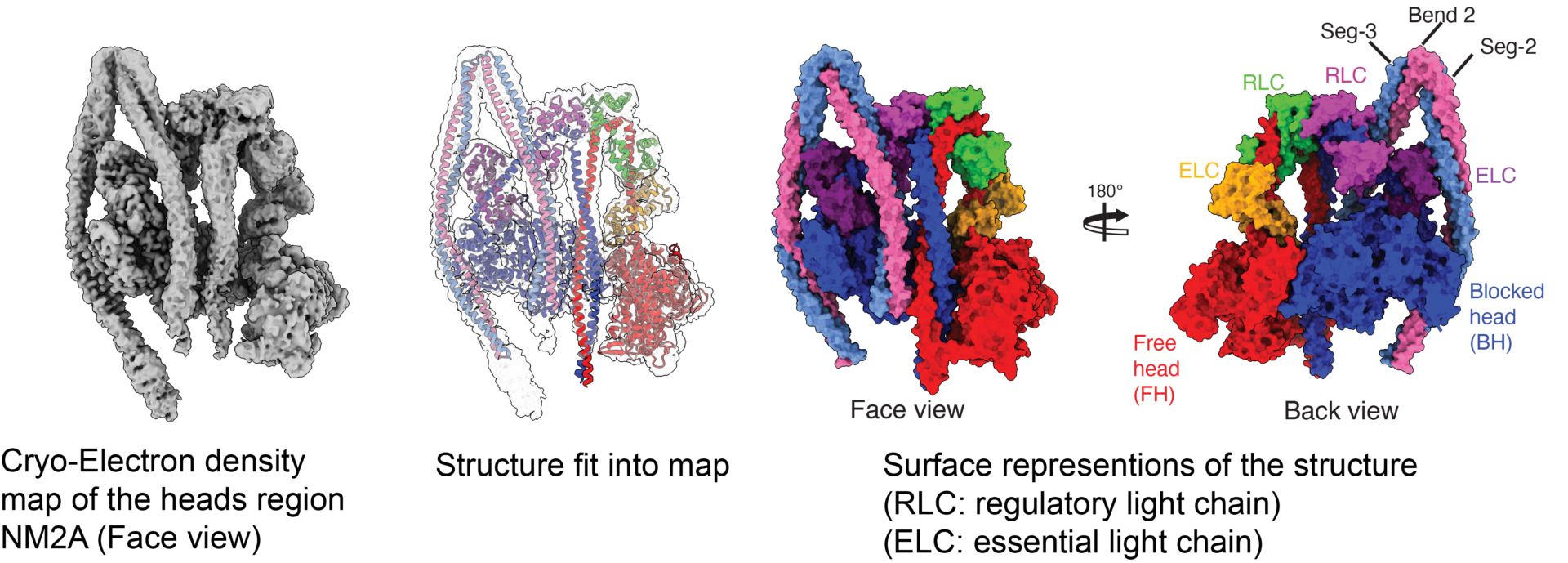
The structure of shutdown platelet myosin.
Our lab has a strong interest in the structure and function of myosin. We want to understand how the activity of myosins is regulated, and how disease mutations affect regulation and function. We have used CryoEM to solve the shutdown structures of smooth and non-muscle myosin 2A (the ‘platelet’ myosin), revealing how these myosins switch off their activity. This has been critical in revealing how mutations in platelet myosin destablise the shutdown state of platelet myosin, leading to bleeding and other disorders. We have determined how specific mutations in β-cardiac myosin likely cause heart and skeletal muscle diseases. We are currently working on ‘unconventional’ myosins (Myosin 5, Myosin 7a) to understand their structure, function and regulation and roles in disease using single particle CryoEM. We are also investigating the organisation of smooth and non-muscle myosins in situ using CryoET.
Super-resolution imaging

dSTORM imaging of a heart muscle myofibril, labelled for ɑ-actinin-2 (Z-disc protein). Colour scheme – depth coded.
We have developed single molecule localisation microscopy approaches to obtain detailed information on protein organisation within the Z-disc, a narrow (~100nm wide) structure found at the ends of muscle sarcomeres. Over 100 proteins are associated with the Z-disc, and yet most seem invisible by CryoET. Single molecule fluorescence imaging, combined with our novel software (PERPL) allows us to pinpoint the organisation of specific proteins within the Z-disc. This approach is facilitated by using Adhirons (small non‑antibody binding proteins) specific for each protein of interest (with the Tomlinson Group @ Leeds). Their small size (~2nm, 10 kDa) means better penetration into dense cytoskeletal regions, a lower linkage error and thus better resolution in our final images.
Current PhD projects include:
- Using imaging (fluorescence, super-resolution, CryoET) of diseased heart tissue to understand the consequences of disease mutations on heart structure and function (BHF funded student),
- Using imaging (Fluorescence, Super-resolution, CryoET) and single-particle CryoEM, to investigate smooth muscle structure and function, and smooth muscle myosin regulation (BBSRC PhD student)
- Building a new super-resolution microscope for fast, high-resolution live cell imaging and expansion microscopy (Nanoram, EU project).