Swinging on "monkey bars": motor proteins caught in the act
The first images of motor proteins in action are published in the journal Nature Communications today.
These proteins are vital to complex life, forming the transport infrastructure that allows different parts of cells to specialise in particular functions. Until now, the way they move has never been directly observed.
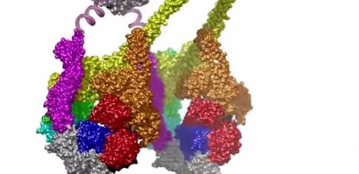
Researchers at the University of Leeds and in Japan used electron microscopes to capture images of the largest type of motor protein, called dynein, during the act of stepping along its molecular track.
Dr Stan Burgess, at the University of Leeds’ School of Molecular and Cellular Biology, who led the research team, said: “Dynein has two identical motors tied together and it moves along a molecular track called a microtubule. It drives itself along the track by alternately grabbing hold of a binding site, executing a power stroke, then letting go, like a person swinging on monkey bars.
“Previously, dynein movement had only been tracked by attaching fluorescent molecules to the proteins and observing the fluorescence using very powerful light microscopes. It was a bit like tracking vehicles from space with GPS. It told us where they were, their speed and for how long they ran, stopped and so on, but we couldn’t see the molecules in action themselves. These are the first images of these vital processes.”
Read the full press release on the Faculty of Biological Sciences website