Macromolecular X-ray Crystallography
 Application

Understanding the biological function of different macromolecules by using X-ray crystallograhy to determine their three dimensional structures. Methods employed include crystallisation, X-ray diffraction of macromolecule crystals, processing of collected diffraction images, model building and refinement. The molecular and structural information obtained could ultimately provide a better understanding of the mechanism of these macromolecules.
Equipment
Protein crystallisation screen liquid handler
- Formulator (FormulatrixÒ)
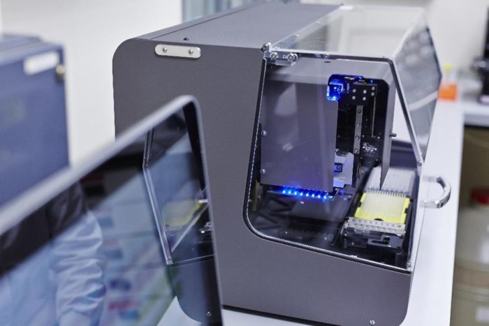
Robotic crystallisation system
- NT8 LCP drop setter (FormulatrixÒ)
- Mosquito Crystal (ttplabtech)
Crystallisation imaging system (FormulatrixÒ)
- Rock Imager 1000 at 20°C with Second Order Nonlinear Imaging of Chiral Crystals (SONICC) imaging.
- Rock Imager 1000 at 4 °C with UV imaging.
- Fluorescence Recovery After Photobleaching (FRAP) imaging to Identify the optimal conditions for Lipidic Cubic Phase crystal growth for membrane protein crystallography.
X-ray diffraction data collection is available at both the Diamond Light Source Ltd, Oxfordshire and the European Synchrotron Radiation Facility, France.
Location
Room 8.116 Astbury Building
Contact
 Dr Chi Trinh
School of Molecular & Cellular Biology
Email: c.h.trinh@leeds.ac.uk
Tel: 0113 343 3023
Further information
https://biologicalsciences.leeds.ac.uk/facilities/doc/x-ray-crystallography-1